

當(dāng)前位置: 首頁 - 產(chǎn)品專區(qū) - 熱銷產(chǎn)品

H3K9/14ac polyclonal antibody
| 貨號 | C15410200-10/C15410200-50 | 售價(元) | 咨詢 |
| 規(guī)格 | 10ug/50ug | CAS號 |
- 產(chǎn)品簡介
- 相關(guān)產(chǎn)品
Polyclonal antibody raised in rabbit against the region of histone H3 containing the acetylated lysines 9 and 14 (H3K9/14ac), using a KLH-conjugated synthetic peptide.
| Lot | A1756D |
|---|---|
| Concentration | 0.81 μg/μl |
| Species reactivity | Human, mouse, P. falciparum, wide range expected. |
| Type | Polyclonal, ChIP grade, ChIP-seq grade |
| Purity | Affinity purified polyclonal antibody. |
| Host | Rabbit |
| Storage Conditions | Store at -20°C; for long storage, store at -80°C. Avoid multiple freeze-thaw cycles. |
| Storage Buffer | PBS containing 0.05% azide and 0.05% ProClin 300. |
| Precautions | This product is for research use only. Not for use in diagnostic or therapeutic procedures. |
| Applications | Suggested dilution | References |
|---|---|---|
| ChIP/ChIP-seq * | 1 μg per IP | Fig 1, 2 |
| CUT&TAG | 1 μg | Fig 3 |
| ELISA | 1:100 | Fig 4 |
| Dot Blotting/Peptide array | 1:20,000/1:2,000 | Fig 5 |
| Western Blotting | 1:500 | Fig 6 |
| Immunofluorescence | 1:200 | Fig 7 |
* Please note that the optimal antibody amount per ChIP should be determined by the end-user. We recommend testing 1-5 μg per IP.
-
Validation data
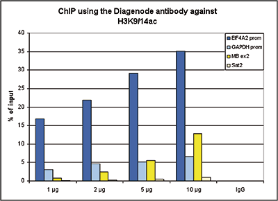
Figure 1. ChIP results obtained with the Diagenode antibody directed against H3K9/14ac
ChIP assays were performed using human HeLa cells, the Diagenode antibody against H3K9/14ac (Cat. No. C15410200) and optimized PCR primer pairs for qPCR. ChIP was performed with the “Auto Histone ChIP-seq” kit on the IP-Star automated system, using sheared chromatin from 1,000,000 cells. A titration consisting of 1, 2, 5 and 10 μg of antibody per ChIP experiment was analyzed. IgG (2 μg/IP) was used as a negative IP control. Quantitative PCR was performed with primers specific for the promoter of the active genes GAPDH and EIF4A2, used as positive controls, and for the coding region of the inactive MB gene and the Sat2 satellite repeat, used as negative controls. Figure 1 shows the recovery, expressed as a % of input (the relative amount of immunoprecipitated DNA compared to input DNA after qPCR analysis).A.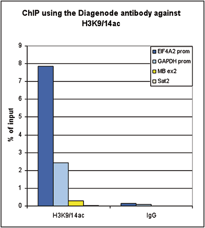
B.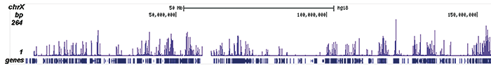
C.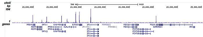
D.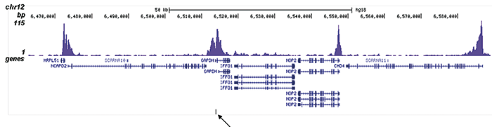
E.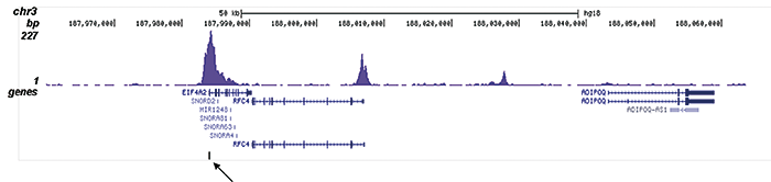
Figure 2. ChIP-seq results obtained with the Diagenode antibody directed against H3K9/14ac
ChIP was performed on sheared chromatin from 100,000 K562 cells using 1 μg the Diagenode antibody against H3K9/14ac (Cat. No. C15410200) with the “iDeal ChIP-seq” kit (Cat. No. C01010051). IgG (1 μg/IP) was used as a negative IP control. The IP’d DNA was analysed by QPCR with optimized PCR primer pairs for the promoters of the active GAPDH and EIF4A2 genes, used as positive control targets, and the coding region of the inactive MB gene and the Sat2 satellite repeat, used as negative control targets (figure 2A). The IP’d DNA was subsequently analysed with an Illumina Genome Analyzer. Library preparation, cluster generation and sequencing were performed according to the manufacturer’s instructions. The 36 bp tags were aligned to the human genome using the ELAND algorithm. Figure 2 shows the peak distribution along the complete sequence and a 1.5 Mb region of the X-chromosome (figure 2B and C) and in two regions surrounding the GAPDH and EIF4A2 positive control genes, respectively (figure 2D and E). The position of the amplicon used for ChIP-qPCR is indicated by an arrow. These results clearly show an enrichment of the H3K9/14 acetylation at the promoters of active genes.A.
B.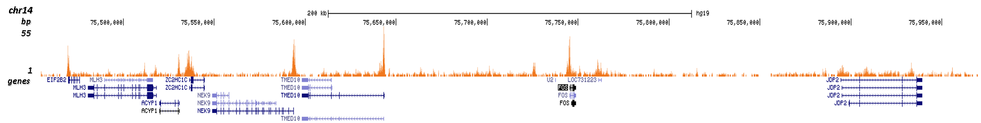
Figure 3. Cut&Tag results obtained with the Diagenode antibody directed against H3K9/14ac
CUT&TAG (Kaya-Okur, H.S., Nat Commun 10, 1930, 2019) was performed on 50,000 K562 cells using 1 μg of the Diagenode antibody against H3K9/14ac (cat. No. C15410200) and the Diagenode pA-Tn5 transposase (C01070001). The libraries were subsequently analysed on an Illumina NextSeq 500 sequencer (2x75 paired-end reads) according to the manufacturer's instructions. The tags were aligned to the human genome (hg19) using the BWA algorithm. Figure 3 shows the peak distribution along the complete sequence of chromosome 7 and in a 500 kb region surrounding the FOS gene on chromosome 14 (figure 3A and B, respectively).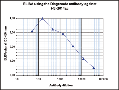
Figure 4. Determination of the antibody titer
To determine the titer of the antibody, an ELISA was performed using a serial dilution of the Diagenode antibody against H3K9/14ac (Cat. No. C15410200). The antigen used was a peptide containing the histone modification of interest. By plotting the absorbance against the antibody dilution (Figure 4), the titer of the antibody was estimated to be 1:4,000.A.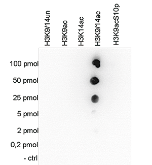
B.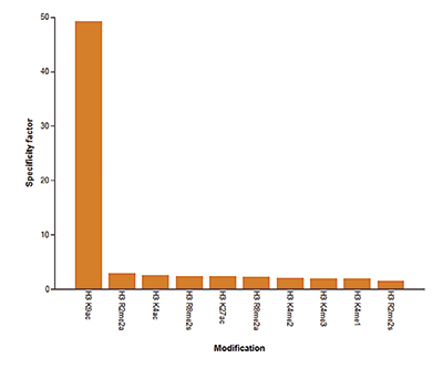
Figure 5. Cross reactivity tests using the Diagenode antibody directed against H3K9/14ac
Figure 5A To test the cross reactivity of the Diagenode antibody against H3K9/14ac (Cat. No. C15410200), a Dot Blot analysis was performed with peptides containing other histone modifications and the unmodified H3K9. One hundred to 0.2 pmol of the respective peptides were spotted on a membrane. The antibody was used at a dilution of 1:20,000. Figure 5A shows a high specificity of the antibody for the modification of interest. Figure 5B The specificity of the antibody was further demonstrated by peptide array analyses on an array containing 384 peptides with different combinations of modifications from histone H3, H4, H2A and H2B. The antibody was used at a dilution of 1:2,000. Figure 5B shows the specificity factor, calculated as the ratio of the average intensity of all spots containing the mark, divided by the average intensity of all spots not containing the mark.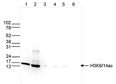
Figure 6. Western blot analysis using the Diagenode antibody directed against H3K9/14ac
Western blot was performed on whole cell (25 μg, lane 1) and histone extracts (15 μg, lane 2) from HeLa cells, and on 1 μg of recombinant histone H2A, H2B, H3 and H4 (lane 3, 4, 5 and 6, respectively) using the Diagenode antibody against H3K9/14ac (Cat. No. C15410200). The antibody was diluted 1:500 in TBS-Tween containing 5% skimmed milk. The position of the protein of interest is indicated on the right, the marker (in kDa) is shown on the left.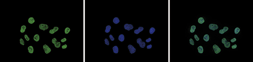
Figure 7. Immunofluorescence using the Diagenode antibody directed against H3K9/14ac
HeLa cells were stained with the Diagenode antibody against H3K9/14ac (Cat. No. C15410200) and with DAPI. Cells were fixed with 4% formaldehyde for 10’ and blocked with PBS/TX-100 containing 5% normal goat serum and 1% BSA. The cells were immunofluorescently labeled with the H3K9/14ac antibody (left) diluted 1:200 in blocking solution followed by an anti-rabbit antibody conjugated to Alexa488. The middle panel shows staining of the nuclei with DAPI. A merge of the two stainings is shown on the right.