

當(dāng)前位置: 首頁(yè) - 產(chǎn)品專區(qū) - 熱銷產(chǎn)品

Pol II monoclonal antibody
| 貨號(hào) | C15200004-10/C15200004-50 | 售價(jià)(元) | 咨詢 |
| 規(guī)格 | 10ug/50ug | CAS號(hào) |
- 產(chǎn)品簡(jiǎn)介
- 相關(guān)產(chǎn)品
Alternative names: POLR2A, RPB1, POLR2, RPOL2
Monoclonal antibody raised in mouse against the YSPTSPS repeat in the B1 subunit of RNA polymerase II.
| Lot | 001-14 |
|---|---|
| Concentration | 1.0 μg/μl |
| Species reactivity | Human, Xenopus, Yeast: positive. Other species: not tested. |
| Type | Monoclonal ChIP grade, ChIP-seq grade |
| Purity | Protein A purified monoclonal antibody. |
| Host | Mouse |
| Storage Conditions | Store at -20°C; for long storage, store at -80°C. Avoid multiple freeze-thaw cycles. |
| Storage Buffer | PBS containing 0.05% azide. |
| Precautions | This product is for research use only. Not for use in diagnostic or therapeutic procedures. |
| Applications | Suggested dilution | References |
|---|---|---|
| ChIP/ChIP-seq * | 1 μg/ChIP | Fig 1, 2 |
| ELISA | 1:3,000 | Fig 3 |
| Western Blotting | 1:1,000 | Fig 4, 5 |
| Immunofluorescence | 1:500 | Fig 6 |
* Please note that the optimal antibody amount per ChIP should be determined by the end-user. We recommend testing 1-5 μg per IP.
-
Validation data
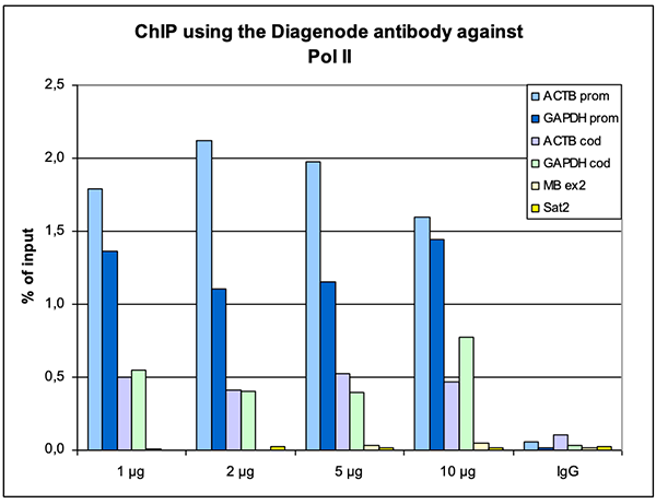
Figure 1. ChIP results obtained with the Diagenode monoclonal antibody directed against Pol II
ChIP assays were performed using human HeLa cells, the Diagenode monoclonal antibody against Pol II (Cat. No. C15200004) and optimized PCR primer pairs for qPCR. ChIP was performed with the "iDeal ChIP-seq" kit (Cat. No. C01010051), using sheared chromatin from 1 million cells. A titration consisting of 1, 2, 5 and 10 μg of antibody per ChIP experiment was analyzed. IgG (2 μg/IP) was used as a negative IP control. Quantitative PCR was performed with primers specific for the promoter and the coding region of the constitutively expressed GAPDH and ACTB genes, used as positive controls, and for exon 2 of the inactive myoglobin (MB) gene and the Sat2 satellite repeat, used as negative controls. Figure 1 shows the recovery, expressed as a % of input (the relative amount of immunoprecipitated DNA compared to input DNA after qPCR analysis).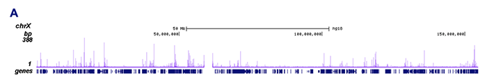
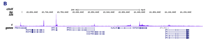
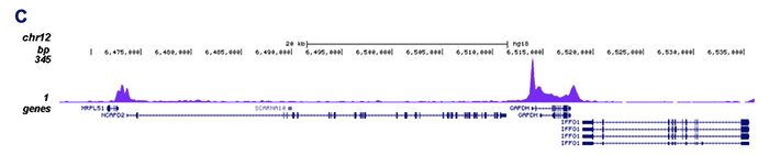
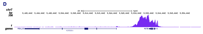
Figure 2. ChIP-seq results obtained with the Diagenode monoclonal antibody directed against Pol II
ChIP was performed on sheared chromatin from 1 million HeLaS3 cells using 1 μg of the Diagenode antibody against Pol II (Cat. No. C15200004) as described above. The IP'd DNA was subsequently analysed on an Illumina Genome Analyzer. Library preparation, cluster generation and sequencing were performed according to the manufacturer's instructions. The 36 bp tags were aligned to the human genome using the ELAND algorithm. Figure 2 shows the peak distribution along the complete sequence and a 400 kb region of the X-chromosome (figure 2A and B, respectively), and in a two genomic regions surrounding the GAPDH and ACTB positive control genes (figure 2C and D).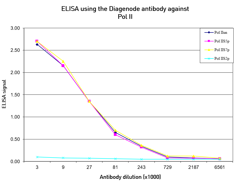
Figure 3. Cross reactivity of the Diagenode monoclonal antibody directed against Pol II
To test the specificity an ELISA was performed using a serial dilution of the Diagenode monoclonal antibody against Pol II (Cat. No. C15200004). The wells were coated with peptides containing the unmodified C-terminal repeat sequence as well as different phosphorylated peptides. Figure 3 shows that the antibody recognizes the unphosphorylated Pol II as well as most phosphorylated forms.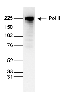
Figure 4. Western blot analysis using the Diagenode monoclonal antibody directed against Pol IINuclear extracts (25 μg) from HeLa cells were analysed by Western blot using the Diagenode monoclonal antibody against Pol II (Cat. No. C15200004) diluted 1:1,000 in TBS-Tween containing 5% skimmed milk. The position of the protein of interest is indicated on the right; the marker (in kDa) is shown on the left.
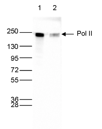
Figure 5. Western blot analysis using the Diagenode monoclonal antibody directed against Pol II
Whole cell extracts (40 μg) from HeLa cells transfected with Pol II siRNA (lane 2) and from an untransfected control (lane 1) were analysed by Western blot using the Diagenode antibody against Pol II (Cat. No. C15200004) diluted 1:1,000 in TBS-Tween containing 5% skimmed milk. The position of the protein of interest is indicated on the right; the marker (in kDa) is shown on the left.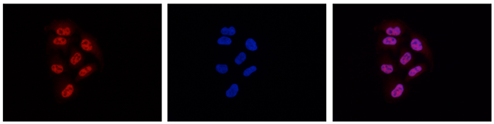
Figure 6. Immunofluorescence using the Diagenode monoclonal antibody directed against Pol II
HeLa cells were stained with the Diagenode antibody against Pol II (Cat. No. C15200004) and with DAPI. Cells were fixed with methanol and blocked with PBS/TX-100 containing 5% normal goat serum and 1% BSA. The cells were immunofluorescently labelled with the Pol II antibody (left) diluted 1:500 in blocking solution followed by an anti-mouse antibody conjugated to Alexa594. The middle panel shows staining of the nuclei with DAPI. A merge of the two stainings is shown on the right.
- 上一頁(yè): 3-methylcytosine (3-mC) Antibody
- 下一頁(yè): HDAC2 monoclonal antibody